

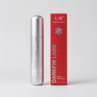

SKIN THAT BOUNCES BACK.
Signaling peptides that survive, and arrive.
L-01 delivers signaling peptides, the messenger molecules skin uses to renew itself, on a plant carrier that keeps them intact and carries them deeper.
SEVEN STUDIES. ONE FORMULA.
From the lab bench to clinical studies, L-01 was tested by independent research labs before it reached you.
WHAT 28 DAYS LOOKS LIKE
A 28-day single-center clinical study. 29 women, ages 25–54, applied the L-01 formula twice daily to the face.1
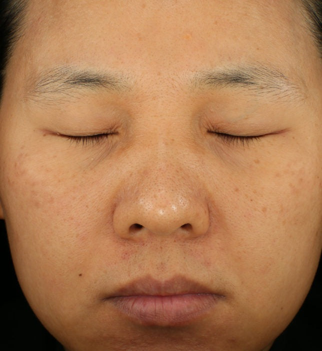
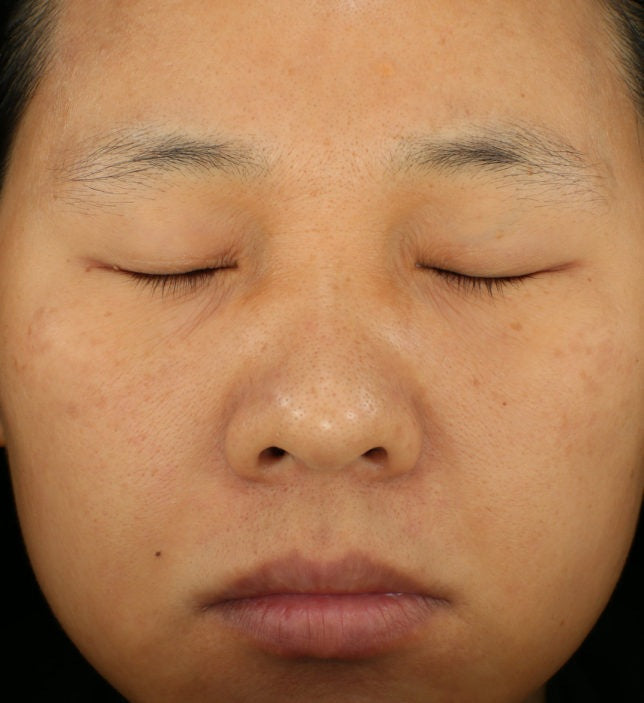
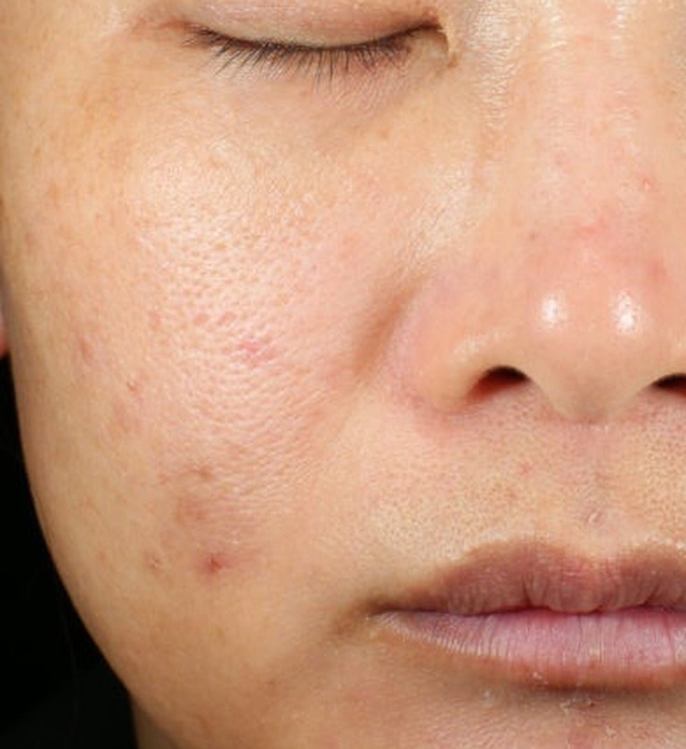
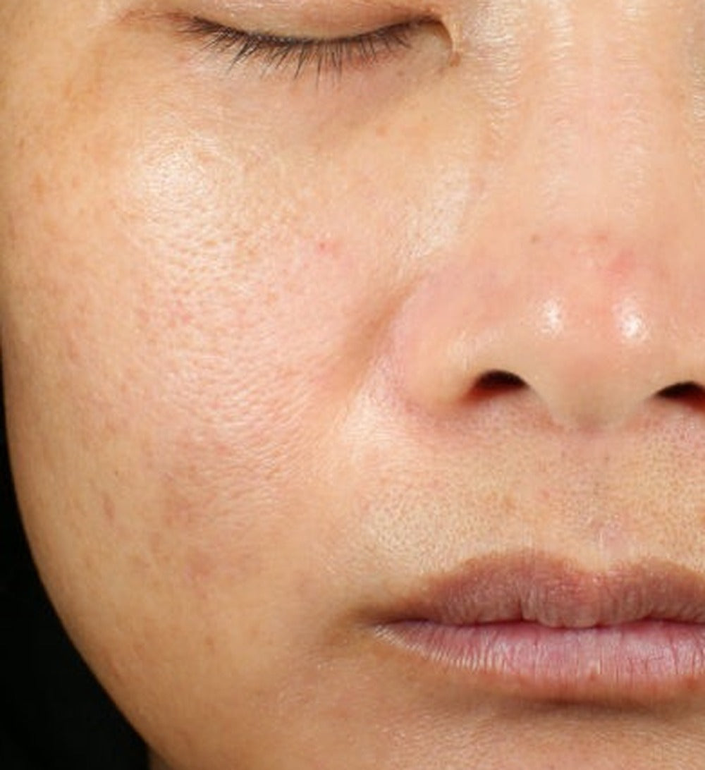
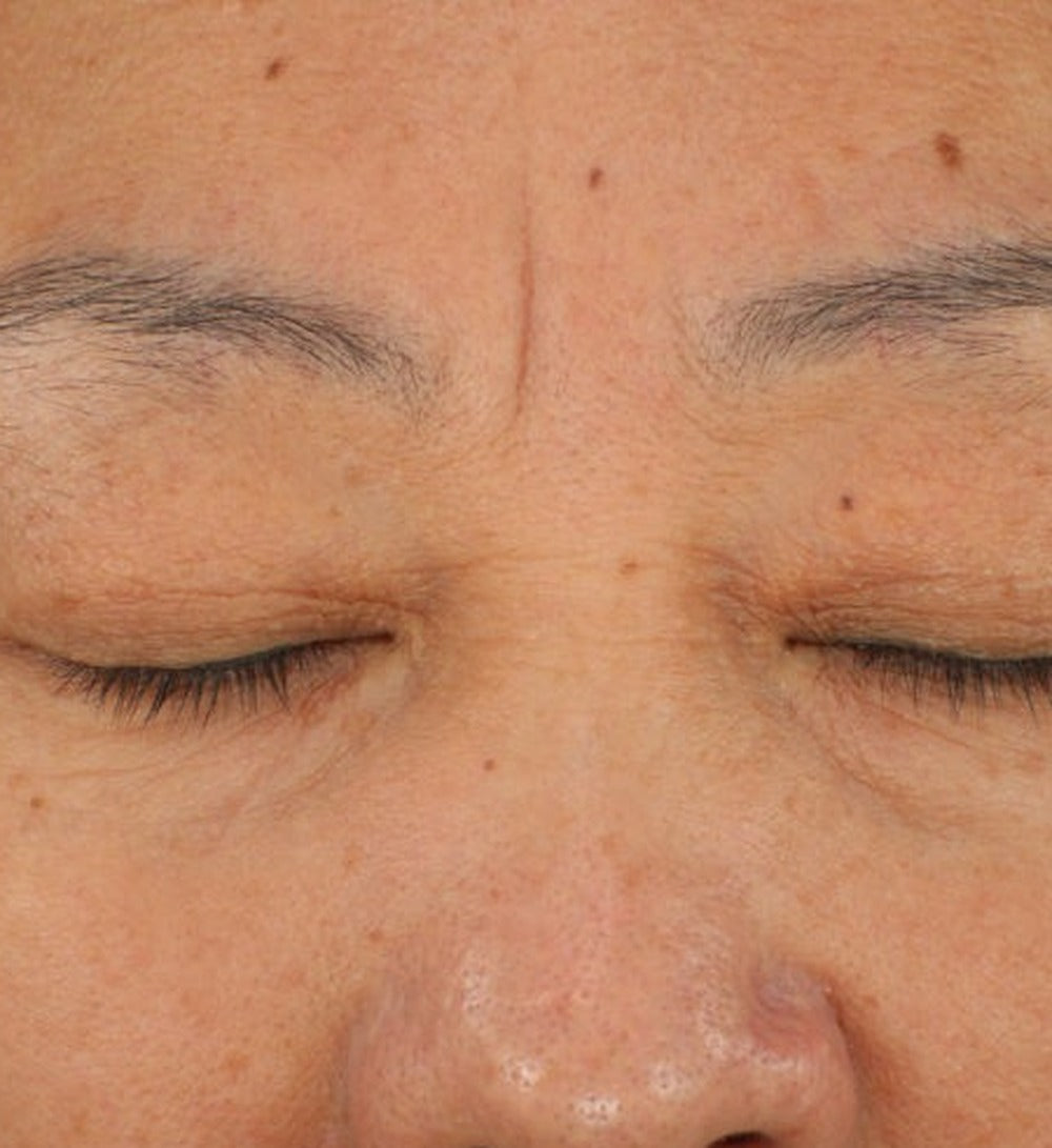
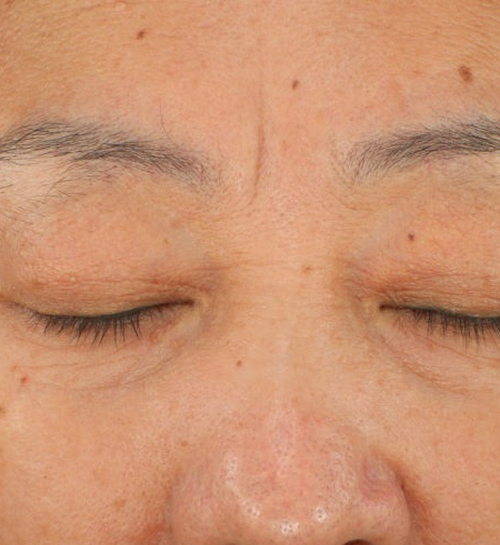
L-01 formula study participants, standardized clinical photography. Individual results vary.1
How it was measured
VISIA-CR · Canfield Scientific. Standardized clinical facial imaging, with the same lighting and position at every visit, so wrinkles can be counted and measured over time.
Tewameter TM Hex · Courage + Khazaka. Measures how much water escapes through the skin (TEWL). Less water loss means a stronger skin barrier.
Dermatologist grading · Standardized grading scales. A dermatologist graded visible facial redness and wrinkles on standardized scales over the study.
Participant efficacy evaluation · 0–9 scale · p<0.001. All 29 participants scored their skin under clinical conditions at day 0, day 14 and day 28.
Dermatologist and instrumental evaluations were performed on a n=10 cohort at baseline and day 28.1
TESTED AGAINST PLACEBO. THEN AGAINST RETINOL.
Two independent clinical studies tested the L-01 signaling-peptide ingredients against a placebo cream and against 0.3% retinol.3
See the full comparison and study images
Across 9 measures, the L-01 ingredients led on 4, tied on 4, and retinol led on 1 (pores).
| Measure | L-01 ingredients | 0.3% retinol |
|---|---|---|
| First significant wrinkle improvement | Day 14 | Day 30 |
| Smoother skin, day 30 | 77% | 59% |
| Brightness | Improved by day 30 | No effect |
| Firmness, elasticity, barrier, dark spots | Improved | Improved |
| Pores | No effect | Improved |


THREE HALLMARKS OF AGING. ONE COMPLEX.
Scientists describe aging as a set of connected “hallmarks”.A In skin, three of them show up as changes you can see. UV light and oxidative stress speed them up.
L-01 was tested on skin cells after UV and oxidative damage. Here is each hallmark, and what L-01 did.5
72% of UV-induced collagen loss prevented5

The problem: Collagen and elastin keep skin firm and springy. With age and UV, they break down. You see: lines, loss of firmness.
With L-01: Protects skin’s support matrix of collagen and elastin, even after UV damage.
−47% senescence marker P165

The problem: Senescent cells stop renewing but don’t go away, and they release inflammatory signals that age the cells around them.A You see: dullness, redness, sensitivity.
With L-01: Fewer signs of cellular aging and calmer inflammatory signals, even after oxidative damage.
+19% faster wound-gap closure5

The problem: Older skin repairs more slowly.F You see: marks that linger, slower recovery.
With L-01: Skin cells close a wound gap faster.
See all 7 markers
7 of 7 markers moved toward the healthy state · all p<0.05. % change vs stressed cells without L-01.
| Marker | Change |
|---|---|
| Collagen I (after UV) | +61% |
| Elastin (after UV) | +17% |
| MMP-1, collagen-cutting enzyme (after UV) | −9% |
| P16, senescence marker (after oxidative stress) | −47% |
| IL-6, inflammatory signal | −14% |
| TNF-α, inflammatory signal | −13% |
| Cell migration, vs untreated | +19% |
No cell toxicity up to about 4× the test dose (≥95% viability).5 Lab results on skin cells (dermal fibroblasts). Our full study is being prepared for peer-reviewed publication.
A DELIVERY BREAKTHROUGH
We bound growth-factor peptides to oleosomes: natural oil bodies from plant seeds. The result: signaling peptides that hold up under heat stress and reach the layer of skin where collagen is made.
After 12 weeks of heat stress, 92% intact. On its own, only 8% survived.4
Measures intact protein (SDS-PAGE), not biological activity. EGF shown.
Without a carrier, it never reached the dermis. With oleosomes, it did.44
Ex vivo lab skin model (EpiDermFT), FGF-2, single time point. Illustration.
BORN FROM STEM CELL SCIENCE

Stem cells are skin’s repair crew: they send the regenerative signals that tell skin to renew. With age, those signals fade.A Our founding insight: aging skin still has the capacity to repair. It’s missing the signals that trigger it.
-
01
Engineered Stem Cells
Our founder and two senior scientists developed and patented Engineered Stem Cells (ESC): synthetic cells that replicate the regenerative signals healthy stem cells produce.
-
02
AI-guided signal selection
Our research parent company, Stemtology, built an AI discovery platform with Microsoft AI Co-Innovation Labs to find signals that work together across skin-aging pathways.
-
03
L-01
Human-identical signaling peptides made through plant-based protein expression, designed to bring stem cell–inspired repair signals to skin.
BUILT FOR SENSITIVE SKIN
Dermatologist-tested on 51 people with sensitive skin. No irritation. No sensitization.2
QUESTIONS, ANSWERED
How soon will I notice a difference?
In our 28-day clinical study, every skin attribute evaluated had improved by about a third at day 14, and by more than half at day 28.1 Individual results vary.
Which results are from the product I’d actually buy?
Results marked ¹ are from the finished L-01 formula. ² is the sensitive-skin safety test. ³ and ⁴ are studies on the L-01 signaling-peptide ingredients. ⁵ is lab work on skin cells.
How does it compare to retinol?
In a 60-day head-to-head study, the L-01 signaling-peptide ingredients improved wrinkle depth by day 14. 0.3% retinol showed significant wrinkle improvement at day 30. Retinol improved pores; the L-01 ingredients did not.3
How do I use it?
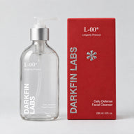
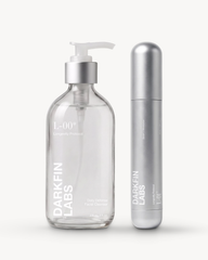
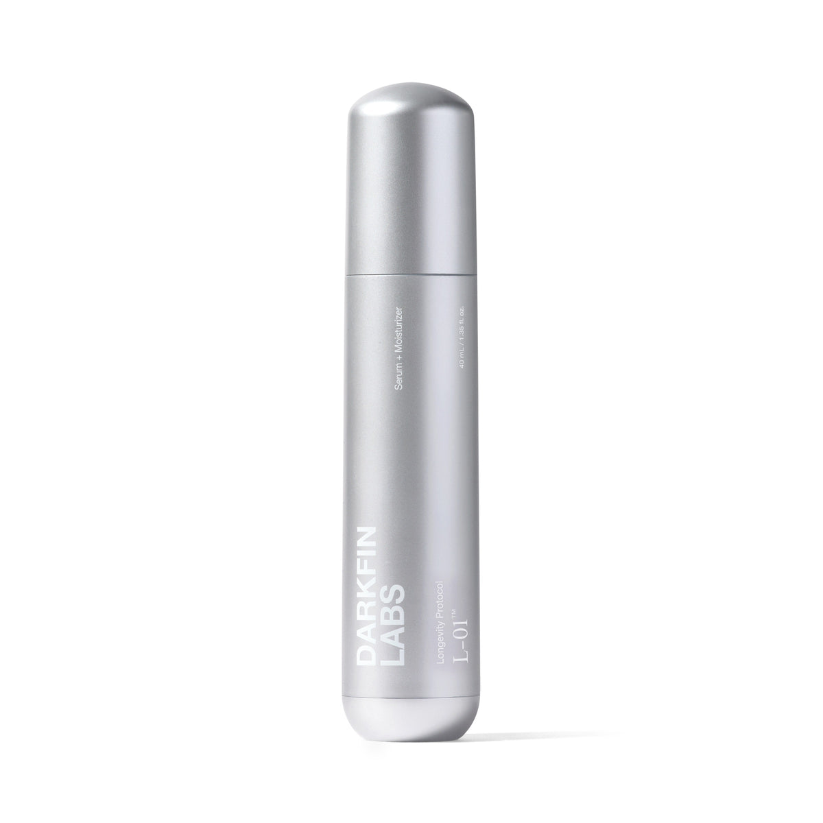
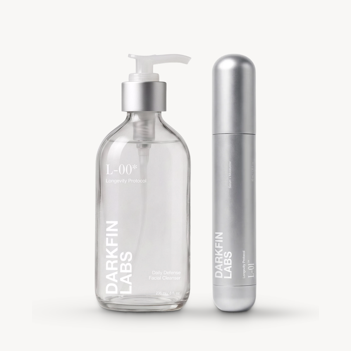
Apply 2–3 pumps to damp skin after cleansing, morning and night. For the full ritual, start with L-00™ Facial Cleanser.
RESULTS YOU CAN FEEL FROM DAY 141
HOW WE REPORT RESULTS
Every number links here. We say what was tested (the finished formula, its ingredients, or skin cells in the lab) and who measured it.
Show study notes & references
- L-01 formula clinical study. A 28-day, single-center clinical study was conducted on 29 females, ages 25–54, with self-reported dull, loose skin lacking elasticity, and visible fine lines and wrinkles. L-01 was applied twice daily to the face. Efficacy and tolerance were evaluated at baseline, day 14, and day 28 by participant efficacy evaluation, dermatologist grading, and instrumental measurement (VISIA-CR imaging, Tewameter TM Hex), with dermatologist and instrumental evaluations performed on a n=10 cohort at baseline and day 28. Participant efficacy evaluation: skin attributes scored on a 0–9 scale under clinical conditions; percentages reflect mean score change from baseline (p<0.001, Wilcoxon signed-rank). Tolerance evaluation: percentage of participants in agreement on a 5-point scale.
- Sensitive-skin safety (RIPT). A dermatologist-tested human repeat insult patch test (RIPT) was conducted on 51 subjects with self-perceived sensitive skin, ages 22–65, Fitzpatrick II–IV. The test material was applied under occlusive patch for nine 48/72-hour induction exposures over three weeks, followed by a 10-day rest and a 48-hour challenge patch read at 48 and 96 hours (ICDRG scale, blinded grading). Under the conditions of the study, there was no indication of a potential to elicit dermal irritation or sensitization. ALS, Torrance, CA; Accession 1277439; 23 Feb – 01 Apr 2026.
- L-01 ingredient clinical studies. Independent clinical studies conducted by Lifeanalytics on the L-01 growth factor oleosome ingredients. Study 1: placebo-controlled, 60 adults, ages 30–65, cream applied twice daily for 28 days; firmness measured by Cutometer R0 at day 14 (+21.8% vs +2.5% placebo, p=0.0003), melanin by Mexameter MX18 at day 28 (−18.0% vs +2.0% placebo, p<0.0001), barrier by TEWL at day 28 (−16.9% vs −5.1% placebo, p=0.0004). Study 2: head-to-head, 44 adults, ages 40–65, growth factor oleosome serum vs 0.3% retinol serum, twice daily for 60 days; maximum wrinkle depth by Antera 3D (−18.6% at day 14, −25.5% at day 30); significant wrinkle improvement at day 14 vs day 30 for retinol. Tested as 1% EGF / 1% FGF-2 growth factor oleosome cream or serum, not as the finished L-01 formula.
- L-01 ingredient lab studies. Stability: in-vitro study, EGF-oleosome fusion vs naked EGF held at 42°C for 12 weeks; intact protein measured by SDS-PAGE band intensity (92% vs 8% of week 0). Measures intact protein, not biological activity. Delivery: ex vivo full-thickness skin model (EpiDermFT), FGF-2-oleosome vs naked FGF-2 at the same dose, Western blot at a single time point; oleosome-bound FGF-2 detected in epidermis and dermis, naked FGF-2 a faint trace in epidermis and none in dermis.
- L-01 complex, skin-cell (in vitro) study. Dermal fibroblasts treated with the L-01 complex at 3.01% (w/w) in an independent laboratory; 3 wells per group. Structure markers (collagen I, elastin, MMP-1) measured after UVA stress; senescence and SASP markers (P16, IL-6, TNF-α) after H₂O₂ oxidative stress; percentages vs stressed cells without L-01. “72% of UV-induced collagen loss prevented” is the share of the stress-induced decrease that L-01 prevented, calculated from group means. Regenerative repair measured by scratch (wound-gap) assay vs untreated cells, no stress model. All 7 markers p<0.05. Cell viability ≥95% up to about 4× the test dose. Lab results on cells. Full study in preparation for peer-reviewed publication.
Scientific references
A. López-Otín C, Blasco MA, Partridge L, Serrano M, Kroemer G. Hallmarks of aging: an expanding universe. Cell. 2023;186(2):243–278.
B. Flament F, Bazin R, Laquieze S, et al. Effect of the sun on visible clinical signs of aging in Caucasian skin. Clin Cosmet Investig Dermatol. 2013;6:221–232.
C. Fisher GJ, Datta SC, Talwar HS, et al. Molecular basis of sun-induced premature skin ageing and retinoid antagonism. Nature. 1996;379:335–339.
D. Ressler S, Bartkova J, Niederegger H, et al. p16INK4A is a robust in vivo biomarker of cellular aging in human skin. Aging Cell. 2006;5(5):379–389.
E. Brincat M, Kabalan S, Studd JW, et al. A study of the decrease of skin collagen content, skin thickness, and bone mass in the postmenopausal woman. Obstet Gynecol. 1987;70(6):840–845.
F. Khalid KA, Nawi AFM, Zulkifli N, et al. Aging and wound healing of the skin: a review of clinical and pathophysiological hallmarks. Life. 2022;12(12):2142.